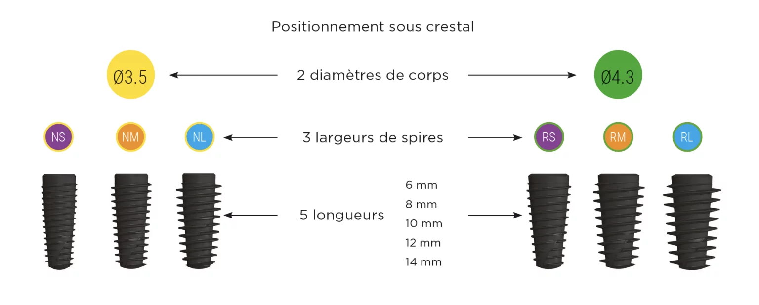
The dental implant designed to preserve bone
etk®, your supplier of ibone E dental implants
Indications for use
– Suitable for all bone densities
– Ideal for poorly vascularised bone
Co-developed with Prof. Hervé TARRAGANO
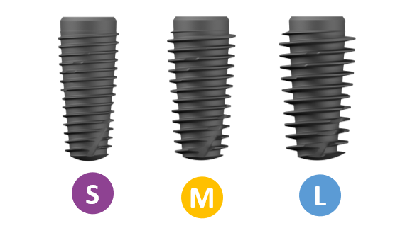
A design intended to guarantee optimal primary stability
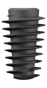
Large threads to leave room for the living
Single and multi-unit connections with the Naturactis, Naturall+ and iBone S dental implant
– Stable implant/prosthetic part assembly
– Precise orientation of prosthetic components
With the same connection for the Naturactis, Naturall+ and iBone S dental implants, it means:
– Streamlining of the inventory of prosthetic parts
– Simplified exchange between the implantologist and its referring dentists
– Simplified exchange between the dental practice and laboratory
A single implant connection for all diameters and all prosthetic platforms:
– Choose whichever gingival platform you want, without worrying about the implant’s diameter. – A simplified treatment plan.
A conical implant body to preserve the bone
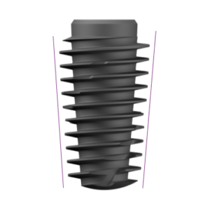
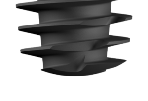
An atraumatic and engaging apex to guarantee ease of insertion while preserving the living
STAE® surface quality, backed by 27 years of clinical experience in surface finish.
– Sandblasting of the titanium oxide to get macroroughness.
– Acid etching to get microroughness.
Your surgery accessories for the iBone E implant
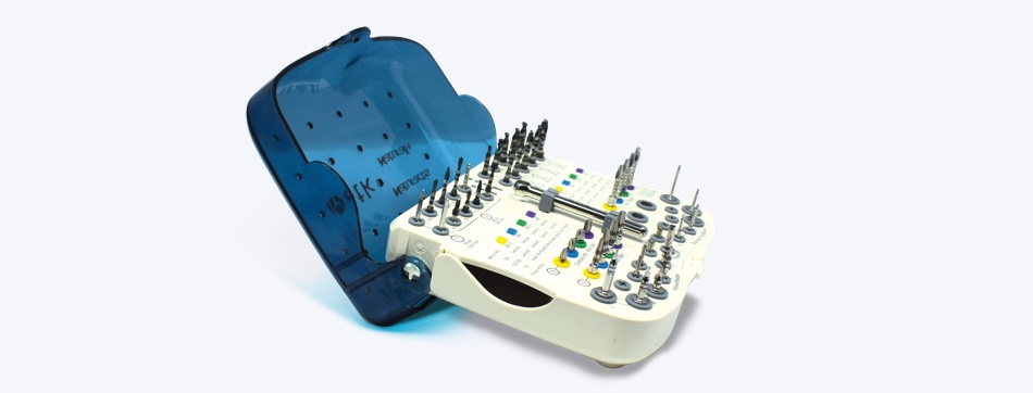
Standard surgical case
– Tiltable for better visibility of the instruments during surgery
– Screen-printed indications for a better understanding of the protocol

Stop kit
– Pick up stops directly from a contra-angle
– Color coding makes identifying stops simple, based on the diameter of the drill used.
– 28 stops for short and long drill bits included in the case Can be autoclave sterilized

Extraction kit
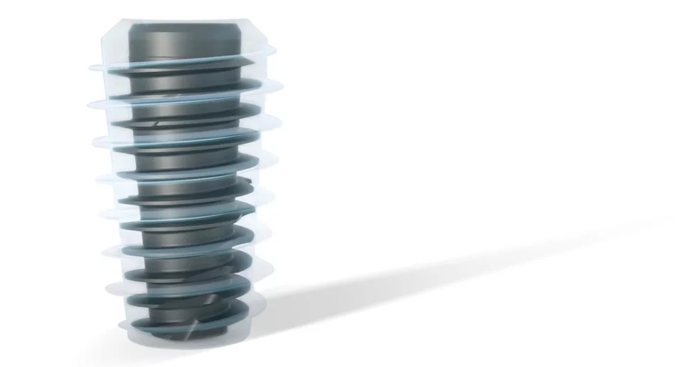
IBone® Dental Implant
As well as offering excellent primary stability, comparable to conventional implants, iBone®, with its reduced body and wide Threads, minimizes the amount of metal in the mouth while providing a more developed contact surface with the bone and space for bone healing. These properties are complemented by a low-torque insertion that minimizes bone heating for increased healing potential and better bone reconstruction.
More than just an implant, iBone® establishes a new protocol in implantology: simple, safe, and accessible, for a less invasive and more conservative result, whatever the implant indication. It is a multi-purpose implant that is more respectful of bone capital and is also a simpler alternative to grafts and sinus lifts.
The IBone® Protocol
In the iBone® protocol, a 'biological bond' is obtained thanks to the empty spaces created at the bone-implant interface. The vascularization of these pockets allows calcium and phosphorus ions present in the blood to be absorbed by the titanium oxide surface of the implant. This calcified afibrillar layer, observed in vitro and in vivo, allows the adhesion of mesenchymal cells, pre-osteoblasts, and osteoblasts, which initiate bone healing.
Less Metal in the Mouth, More Peri-Implant Bone
In the iBone® protocol, priority is given to the bone and the blood clot. The implant's dimensions are reduced as much as possible to create a healing space between the drill hole and the implant. This healing space fills with blood and then with new bone.
The iBone® implant reduces the volume of metal in the bone by up to 34% compared with a conventional implant, allowing the bone to express itself.
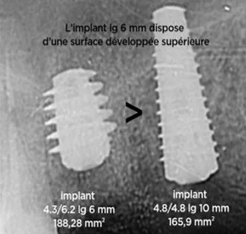
More bone-implant contact surface
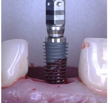
Gentle installation
Excellent primary stability
The average ISQ measured at implant insertion was 73.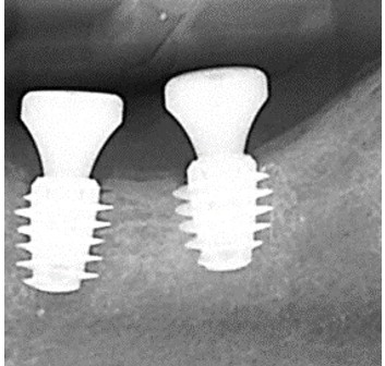
Simplified Surgery
IBone® vs a Conventional Implant
| iBone® implant | Classic implant* | |
| Metal Volume* | 70,91 mm3 | 106,5 mm3 |
| Developed surface | 331,82 mm2 | 211,57 mm2 |
| Torque consumed during installation in D2/D3 bone** | 52,5 N.cm | 80 N.cm |
| Primary stability in D2/D3 (ISQ)** bone | 77.16 | 76 |
Intuitive, Simple and Guided Protocol
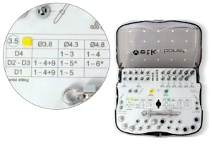
Table of protocols
Numbered drills
Illustration of instruments
Color codes for different diameters
Graduated scale
The perfect osmosis between dentist and prosthetist
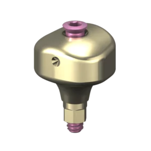
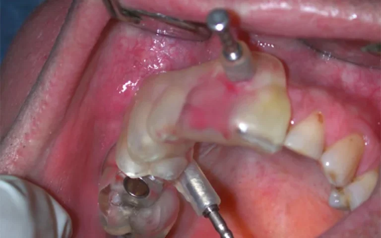
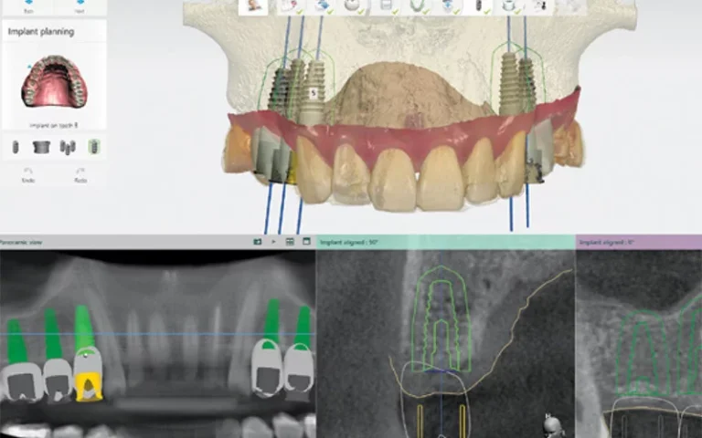
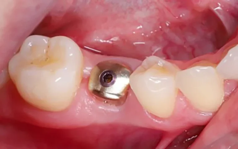
Choose your emergence profile with the iPhysio anatomical abutment corresponding to the tooth. Once fixed in the mouth, it will allow the tissues to develop until the final prosthesis. The impression and a provisional are made on this abutment, without dismantling. The digital impression automatically identifies the abutment to the laboratory, giving it the required emergence profile.
IBone® - Iphysio®: The 100% Bio-Friendly Combo
This unique anatomical part can be used as a healing abutment, a scanbody and a temporary tooth support. It enables the natural emergence profile to be retained throughout treatment without dismantling (no destruction of the mucosal attachment and retention of the emergence profile throughout treatment).
Guided from A to Z, iBone® and iPhysio® constitute the most conservative and codified protocol in implantology.
Optimization Of Implantation In The Posterior Mandibular Sector
Dr Bertrand Hervé, a dental surgeon practicing in Coutances, has been installing iBone® implants for around 3 years. The practitioner illustrates the potential of this new implant design through a clinical case. Two iBone® implants are placed in the mandibular posterior sector to create an implant-supported bridge. This is a standard indication of the iBone® range. In other more constrained areas such as the sub-sinus area, iBone® can avoid complex surgeries or grafts.
Our Reliable And Proven Manufacturing Processes

Certified Surface Finish
30 years of clinical experience Microsandblasting with titanium oxide. Etched with nitric and hydrofluoric acids.

Precise, Watertight, Validated Connection
15 years of clinical experience Internal hexagonal tapered connection. A single prosthetic connection common to all implant diameters.

Our Quality Guarantees
100% French implants Implants guaranteed for life Prosthetic part guaranteed for 10 years CE and ISO 12485 standard
Frequently Asked Questions
1) Allow 2 mm between the implant and the adjacent natural teeth.
2) Allow 3 mm between two adjacent implants.
In the vestibulo-palatine-lingual direction:
1) If possible, leave 1.5 to 2 mm of bone around the vestibular, palatal, and lingual surfaces.


